Teaching NeuroImages: Persistent anterograde amnesia due to sequential, bilateral vascular damage to the Papez circuit
Summary
- 62yo M
- c/c
- presented to emergency dep
- acute impairment of anterograde memory
- Ex
- CT head
- acute changes(–)
- MRI
- acute infact in Lt pos hippo
- nonacute lacunar stroke in Rt ant thalamus
- memory test
- sever deficits in
- verbal
- visual
- sever deficits in
- CT head
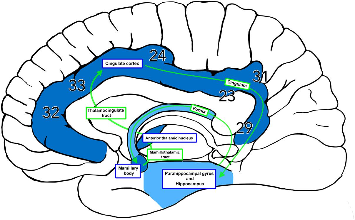
Papez circuit:
- hippocampal formations
- mammillary bodies
- anterior thalamic nuclei
- cingulate and parahippocampal cortices
- their connections via fornix and mammillothalamic tracts
collateral branches of
- PCA
- anterior choroidal A
Further
- Papez circuit (Weininger, Joshua 2019)
- hippocampal formations
- fornix
- mammillary bodies
- mammillothalamic tracts
- anterior thalamic nuclei
- cingulate
- parahippocampal cortices

Terminology
Original
A 62-year-old previously cognitively intact man presented to the emergency department with acute impairment of anterograde memory (ability to form new memories).1 Head CT did not show acute changes; however, amnesia persisted. MRI disclosed an acute infarct in the left posterior hippocampus and a nonacute lacunar stroke in the right anterior thalamus (figure). Memory testing showed severe deficits in verbal and visual memory, persisting after 2 months. Bilateral lesions located anywhere in the Papez circuit (hippocampal formations, mammillary bodies, anterior thalamic nuclei, cingulate and parahippocampal cortices, and their connections via fornix and mammillothalamic tracts) may be implicated in marked, persistent anterograde memory deficits.1,2 The hippocampus receives its blood supply from collateral branches of the posterior cerebral artery and the anterior choroidal artery.3 The anterior thalamus is supplied by the tuberothalamic artery.4
Figure
Acute infarct involving the left hippocampus and nonacute lacunar infarct in the right anterior thalamic nucleus
CT performed in the emergency department with a nonacute right thalamic lacune (A, arrow). Axial diffusion-weighted imaging images show hyperintensity in the left posterior hippocampus (B and C, arrows), signaling diffusion restriction. Coronal T2-weighted (D, arrow) and axial fluid-attenuated inversion recovery (FLAIR) (E, arrow) images depict hyperintensity in the left posterior hippocampus. Axial FLAIR (F, arrow) image confirms a large lacunar infarct in the right anterior thalamic nucleus. L = left; R = right.

References
- Gliebus GP. Memory dysfunction. Continuum (Minneap Minn) 2018;24:727–744.
- Aggleton JP, Pralus A, Nelson AJ, Hornberger M. Thalamic pathology and memory loss in early Alzheimer's disease: moving the focus from the medial temporal lobe to Papez circuit. Brain 2016;139:1877–1890.
- Tatu L, Vuillier F. Structure and vascularization of the human hippocampus. Front Neurol Neurosci 2014;34:18–25.
- Schmahmann JD. Vascular syndromes of the thalamus. Stroke 2003;34:2264–2278.